DIRECT ANSWER
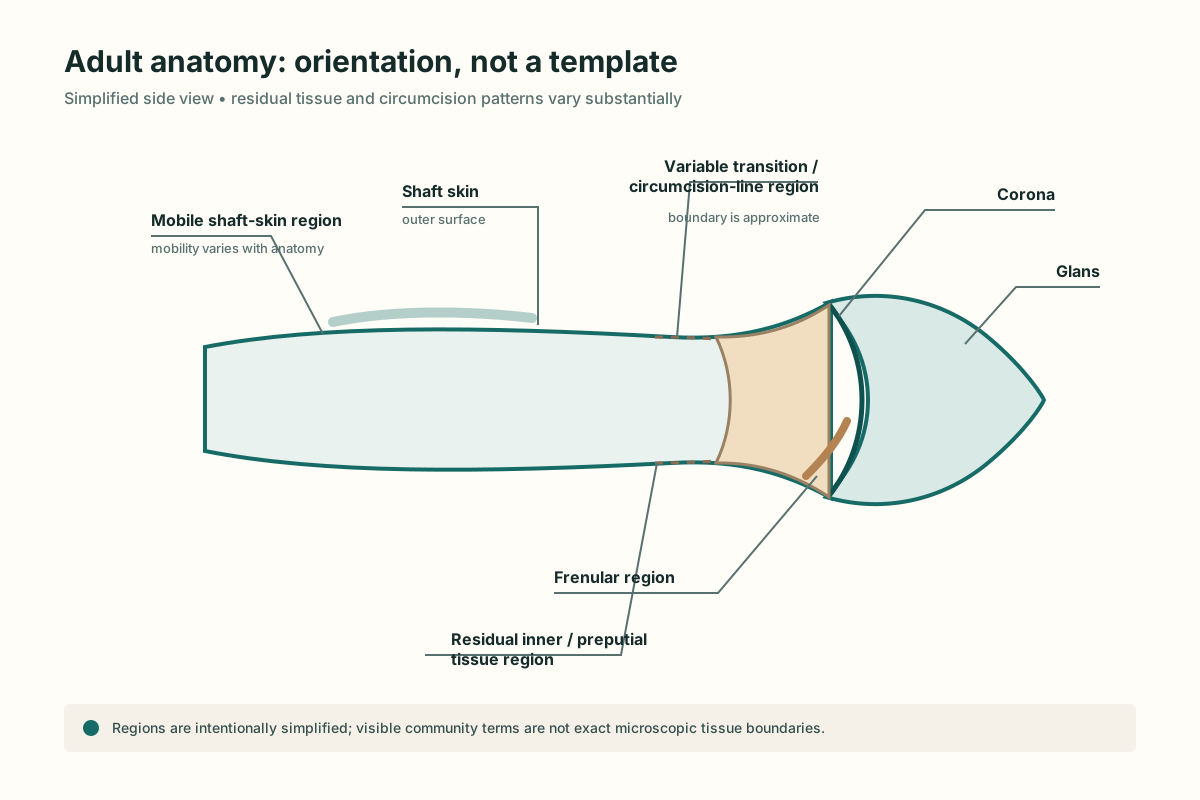
Restoration works with the tissues that remain after circumcision. Relevant structures include mobile shaft skin, residual preputial tissue, the glans and coronal region, any residual frenular tissue, superficial connective tissue, blood vessels, and nerves. Their amount and arrangement vary substantially.
EVIDENCE SUMMARY
How strong and direct is the evidence?
Key landmarks
Shaft skin
A mobile covering over deeper penile structures. Its looseness, hair distribution, scar position, and available movement vary.
Residual preputial tissue
Tissue remaining closer to the glans after circumcision. Restorers often call this “inner skin,” although that is community shorthand.
Glans and corona
The glans is the distal structure; the corona is its rim. Increased coverage changes their surface environment but does not make the covering identical to the removed foreskin.
Frenular region
The ventral region where the frenulum originally connected prepuce and glans. What remains varies markedly.
Mobility and underlying tissue
Penile skin moves relative to deeper structures. Superficial smooth muscle and connective-tissue components commonly discussed under the term dartos contribute to skin behavior, while vessels and nerves support living tissue. Mechanical loading therefore involves more than an isolated surface sheet.
Human studies map development and sensory innervation of the prepuce [1] [2] and describe neural distribution in penile tissues [3]. These studies do not measure restoration outcomes.
Three levels of certainty
Established anatomy
Named structures, tissue layers, vascularity, and innervation can be studied directly in human anatomy and histology.
Community terminology
Terms such as inner skin, outer skin, rollover, and coverage describe practical observations rather than validated anatomical classifications.
Restoration inference
General expansion biology makes growth plausible, but the microscopic organization and sensory outcome of expanded penile tissue are not well characterized.
Why anatomy matters for safety
Clamping, gripping, adhesive contact, or concentrated pressure can affect skin, circulation, and sensation. Fit must account for individual geometry. Numbness, coldness, marked color change, persistent swelling, open skin, or pain are not useful growth targets.