DIRECT ANSWER
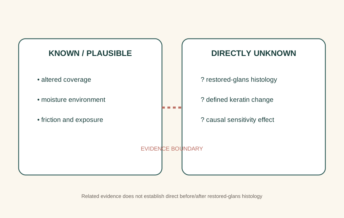
In restoration communities, “dekeratinization” usually describes a glans that appears or feels smoother, less dry, or less exposed after sustained coverage. A changed surface environment is plausible, but direct pre/post histology of restored glans tissue was not located. The term should not be presented as a clinically proven restoration outcome.
EVIDENCE SUMMARY
How strong and direct is the evidence?
Keratin and the stratum corneum
The epidermis matures toward an outer barrier called the stratum corneum. Its flattened, keratin-associated cells and lipids help limit water loss and protect underlying tissue. “Keratinization” can refer broadly to epithelial differentiation or, more narrowly in a study, to measurements of a cornified layer.
“Dekeratinization” is less precise in community use. It may describe visible texture, moisture, shedding, comfort, or an assumed reduction in keratin thickness. Those observations are not interchangeable.
A surface that looks shinier or feels different has changed in some observable way. Demonstrating histological dekeratinization would require tissue sampling or another validated measure of the relevant microscopic feature.
Why coverage could change the surface environment
An exposed glans experiences air, clothing contact, washing, movement, and friction. Sustained tissue coverage could alter moisture retention, direct abrasion, temperature, and exposure. It is reasonable to infer that these conditions may affect surface appearance or feel.
That inference does not specify which microscopic layer changes, by how much, how consistently, or whether a change causes a sensory outcome. Moisture alone can alter appearance and friction without proving a lasting histological transformation.
What the foreskin keratin study measured
Dinh and colleagues sampled inner, outer, and frenular-band foreskin tissue from 19 adults undergoing elective circumcision and measured keratin thickness using histological methods in two laboratories [1]. The study reported substantial variation and did not detect a significant overall difference among the sampled foreskin regions.
This is useful human histology. It challenges simple claims that one foreskin surface always has a much thinner keratin layer. But it examined foreskin tissue—not glans tissue before and after restoration.
Why it cannot answer the restoration question
The tissue, comparison, and timepoint are different. A cross-sectional comparison of foreskin regions cannot demonstrate what sustained restored coverage does to an adult glans.
What microbiota research contributes
Research reviewed by Onywera and colleagues describes associations between circumcision status, penile surface environments, microbial communities, and selected infections [2]. This supports the general point that covered and exposed environments can differ.
Microbial composition is not keratin thickness. Circumcision-status comparisons are not restoration interventions. The review therefore offers surface-environment background, not proof of dekeratinization after restoration.
Visible change, histology, and sensation are separate outcomes
| Observation | What can support it | What remains unproven |
|---|---|---|
| Smoother or more moist appearance | Consistent observation under similar conditions | The microscopic cause |
| Changed friction or comfort | Personal report or validated patient-reported measure | Universal effect or neural mechanism |
| Reduced stratum-corneum thickness | Direct histology or validated tissue measurement | Not established by appearance alone |
| Changed sensitivity | Defined sensory or sexual outcome testing | Not established by keratin or microbiota evidence |
What direct evidence would require
A stronger study would define the restoration exposure, document coverage conditions, examine the same participants before and after sustained coverage, use an appropriate validated method to assess glans histology or barrier characteristics, and separately measure any sensory or comfort outcomes. It would also report confounders such as skin conditions, topical products, hygiene practices, friction, and adherence.
No such direct pre/post restored-glans histology was located for this evidence review.
Use specific observations instead of one overloaded term
More precise language improves both personal notes and research. “The surface appeared less dry under the same conditions,” “clothing friction felt different,” and “a laboratory measurement found a change in stratum-corneum thickness” are three distinct statements. Only the last is direct histological or biophysical evidence.
Specific wording also prevents an observed surface change from being turned into a guaranteed chain of dekeratinization, increased nerve exposure, and improved pleasure. Each step would require its own appropriate evidence.
What we know—and what we do not
What we know
- The stratum corneum is part of the epithelial barrier.
- Coverage plausibly changes moisture, friction, and exposure.
- Foreskin keratin thickness varies within and between people.
- Penile surface environments differ across circumcision-status contexts.
- Some restorers report visible or experiential surface changes.
What we do not know
- Whether restored-glans keratin thickness changes
- The magnitude or timing of any histological change
- Whether visible changes persist under all conditions
- Whether a surface change causes sensory improvement
- Whether retention or a particular method changes the outcome
Language that fits the evidence
It is reasonable to say that sustained coverage may change the glans surface environment and that some people report a different appearance or feel. It is not reasonable to state that restoration has been clinically proven to dekeratinize the glans or that dekeratinization restores sensitivity. The first wording identifies an inference and observation; the second invents a demonstrated biological and causal chain.